THOUSANDS OF FREE BLOGGER TEMPLATES

Friday, November 11, 2011

DNA-RNA-Protein

DNA carries the genetic information of a cell and consists of thousands of genes. Each gene serves as a recipe on how to build a protein molecule. Proteins perform important tasks for the cell functions or serve as building blocks. The flow of information from the genes determines the protein composition and thereby the functions of the cell.

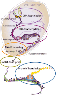
The DNA is situated in the nucleus, organized into chromosomes. Every cell must contain the genetic information and the DNA is therefore duplicated before a cell divides (replication). When proteins are needed, the corresponding genes are transcribed into RNA (transcription). The RNA is first processed so that non-coding parts are removed (processing) and is then transported out of the nucleus (transport). Outside the nucleus, the proteins are built based upon the code in the RNA (translation).

The document has two levels, basic and advanced. This page is an introduction to both levels. You start at the basic level, then you can advance if you want more and deeper information.

Monday, September 12, 2011

kathmandu

The rubber misrepresents an aircraft within an expensive diet.

Monday, August 29, 2011

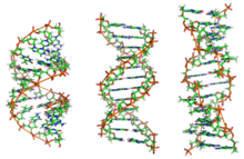
Alternate DNA structures


DNA exists in many possible conformations that include A-DNA, B-DNA, and Z-DNA forms, although, only B-D

NA and Z-DNA have been directly observed in functional organisms.The conformation that DNA adopts depends on the hydration level, DNA sequence, the amount and direction of supercoiling, chemical modifications of the bases, the type and concentration of metalions, as well as the presence of polyamines in solution.

The first published reports of A-DNA X-ray diffraction patterns— and also B-DNA used analyses based on Patterson transforms that provided only a limited amount of structural information for oriented fibers of DNA.An alternate analysis was then proposed by Wilkins et al., in 1953, for the in vivo B-DNA X-ray diffraction/scattering patterns of highly hydrated DNA fibers in terms of squares of Bessel functions. In the same journal, James D. Watson and Francis Crick presented their molecular modeling analysis of the DNA X-ray diffraction patterns to suggest that the structure was a double-helix.

Although the `B-DNA form' is most common under the conditions found in cells, it is not a well-defined conformation but a family of related DNA conformations that occur at the high hydration levels present in living cells. Their corresponding X-ray diffraction and scattering patterns are characteristic of molecular paracrystals with a significant degree of disorder.

Compared to B-DNA, the A-DNA form is a wider right-handed spiral, with a shallow, wide minor groove and a narrower, deeper major groove. The A form occurs under non-physiological conditions in partially dehydrated samples of DNA, while in the cell it may be produced in hybrid pairings of DNA and RNA strands, as well as in enzyme-DNA complexes.Segments of DNA where the bases have been chemically modified by methylation may undergo a larger change in conformation and adopt the Z form. Here, the strands turn about the helical axis in aleft-handed spiral, the opposite of the more common B form. These unusual structures can be recognized by specific Z-DNA binding proteins and may be involved in the regulation of transcription.

Properties of DNA

DNA is a long polymer made from repeating units called nucleotides. As first discovered by James D. Watson and Francis Crick, the structure of DNA of all species comprises two helical chains each coiled round the same axis, and each with a pitch of 34 Ångströms (3.4 nanometres) and a radius of 10 Ångströms(1.0 nanometres).According to another study, when measured in a particular solution, the DNA chain measured 22 to 26 Ångströms wide (2.2 to 2.6 nanometres), and one nucleotide unit measured 3.3 Å (0.33 nm) long.Although each individual repeating unit is very small, DNA polymers can be very large molecules containing millions of nucleotides. For instance, the largest human chromosome, chromosome number 1, is approximately 220 million base pairslong.

In living organisms DNA does not usually exist as a single molecule, but instead as a pair of molecules that are held tightly together. These two long strands entwine like vines, in the shape of a double helix. The nucleotide repeats contain both the segment of the backbone of the molecule, which holds the chain together, and a nucleobase, which interacts with the other DNA strand in the helix. A nucleobase linked to a sugar is called a nucleoside and a base linked to a sugar and one or more phosphate groups is called a nucleotide. Polymers comprising multiple linked nucleotides (as in DNA) is called a polynucleotide.

The backbone of the DNA strand is made from alternating phosphate and sugarresidues. The sugar in DNA is 2-deoxyribose, which is a pentose (five-carbon) sugar. The sugars are joined together by phosphate groups that form phosphodiester bonds between the third and fifth carbon atoms of adjacent sugar rings. These asymmetric bonds mean a strand of DNA has a direction. In a double helix the direction of the nucleotides in one strand is opposite to their direction in the other strand: the strands are antiparallel. The asymmetric ends of DNA strands are called the 5′ (five prime) and 3′ (three prime) ends, with the 5' end having a terminal phosphate group and the 3' end a terminal hydroxyl group. One major difference between DNA and RNA is the sugar, with the 2-deoxyribose in DNA being replaced by the alternative pentose sugar ribose in RNA.
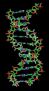
A section of DNA. The bases lie horizontally between the two spiraling strands.Animated version at File:DNA orbit animated.gif.

The DNA double helix is stabilized primarily by two forces: hydrogen bonds between nucleotides and base-stacking interactions among the aromatic nucleobases. In the aqueous environment of the cell, the conjugated π bonds of nucleotide bases align perpendicular to the axis of the DNA molecule, minimizing their interaction with the solvation shell and therefore, the Gibbs free energy. The four bases found in DNA are adenine (abbreviated A), cytosine (C), guanine (G) and thymine (T). These four bases are attached to the sugar/phosphate to form the complete nucleotide, as shown for adenosine monophosphate.

The nucleobases are classified into two types: the purines, A and G, being fused five- and six-memberedheterocyclic compounds, and the pyrimidines, the six-membered rings C and T. A fifth pyrimidine nucleobase, uracil (U), usually takes the place of thymine in RNA and differs from thymine by lacking amethyl group on its ring. Uracil is not usually found in DNA, occurring only as a breakdown product of cytosine. In addition to RNA and DNA a large number of artificial nucleic acid analogues have also been created to study the proprieties of nucleic acids, or for use in biotechnology.
Tuesday, June 28, 2011

DNA, RNA, and Protein: Life at its simplest

DNA: Deoxyribonucleic acid. The double-stranded chemical instruction manual for everything a plant or animal does: grow, divide, even when and how to die. Very stable, has error detection and repair mechanisms. Stays in the cell nucleus. Can make good copies of itself.

RNA: Ribonucleic acid. Single-stranded where DNA is double-stranded, messenger RNA carries single pages of instructions out of the nucleus to places they're needed throughout the cell. No error detection or repair; makes flawed copies of itself. Evolves ten times faster than DNATransfer RNA helps translate the mRNA message into chains of amino acids in the ribosomes.

[Diagram of RNA vs. DNA: chemical structure and composition]


Base: a building block of DNA and RNA. There are five different bases: Adenine, Thymine, Guanine, Cytosine, and Uracil (which is found only in RNA and replaces Thymine in DNA).


Ribosomes: Message centers throughout the cell where the information from DNA arrives in the form of messenger RNA. The RNA message gets translated into a form the ribosome can understand and tells it which protein building blocks it needs and in what order to assemble them. Ribosomal RNA helps the translation go smoothly.

Amino acids: Polypeptide (protein) building blocks.

Polypeptides: chains of amino acids. Proteins are made up of several or many polypeptides.

Proteins: Chemicals that make up cell and organ structure and carry out reactions throughout the body, from breaking down food to fighting off disease.


DNA is transcribed into mRNA which is translated into amino acids.

This is

DNA-RNA-Protein


Introduction

DNA carries the genetic information of a cell and consists of thousands of genes. Each gene serves as a recipe on how to build a protein molecule. Proteins perform important tasks for the cell functions or serve as building blocks. The flow of information from the genes determines the protein composition and thereby the functions of the cell.
The DNA is situated in the nucleus, organized into chromosomes. Every cell must contain the genetic information and the DNA is therefore duplicated before a cell divides (replication). When proteins are needed, the corresponding genes are transcribed into RNA (transcription). The RNA is first processed so that non-coding parts are removed (processing) and is then transported out of the nucleus (transport). Outside the nucleus, the proteins are built based upon the code in the RNA (translation).
The document has two levels, basic and advanced. This page is an introduction to both levels. You start at the basic level, then you can advance if you want more and deeper information.


Saturday, June 11, 2011

Southern Blotting


This technique, named after its inventor E.M. Southern, is a way of combining restriction enzyme and hybridization information. In Southern blotting, a DNA is digested with a restriction enzyme and the fragments are separated by gel electrophoresis. The DNA fragments are denatured by soaking the gel in base and then are transferred to a piece of nitrocellulose filter paper by capillary action. The result is like a “contact print” of the gel; each fragment is at a position on the paper that corresponds exactly to its position in the gel. Then the filter paper is mixed with a radioactively labeled single-stranded “probe” DNA or RNA. The probe DNA hybridizes to the complementary single-stranded DNA fragments on the filter paper. The blot is exposed to X-ray film and the resulting autoradiograph shows the positions of the complementary fragments of DNA. This information physically maps the regions of a large DNA that are complementary to a probe. See Figure 1 . 


Molecular Cloning


DNA replication involves the copying of each strand of the double helix to give a pair of daughter strands. Replication begins at a specific sequence, called the origin. After initiation begins at an origin sequence, all sequences are replicated no matter what their information. This principle leads to the idea of molecular cloning, or recombinant DNA. Cloning enables the production of a single DNA sequence in large quantities.
A recombinant DNA consists of two parts: a vector and the passenger sequences. Vectors supply replication functions—the origin sequences to the recombinant DNA molecule. After it joins to a vector, any passenger sequence can be replicated. The process of joining the vector and passenger DNAs is called ligationDNA ligasecarries out ligation by using ATP energy to make the phosphodiester bond between the vector and passenger. If the vector and passenger DNA fragments are produced by the action of the same restriction endonuclease, they will join by base-pairing. After they are ligated to a vector, it is possible to make an essentially unlimited amount of the passenger sequence.

Plasmid vectors

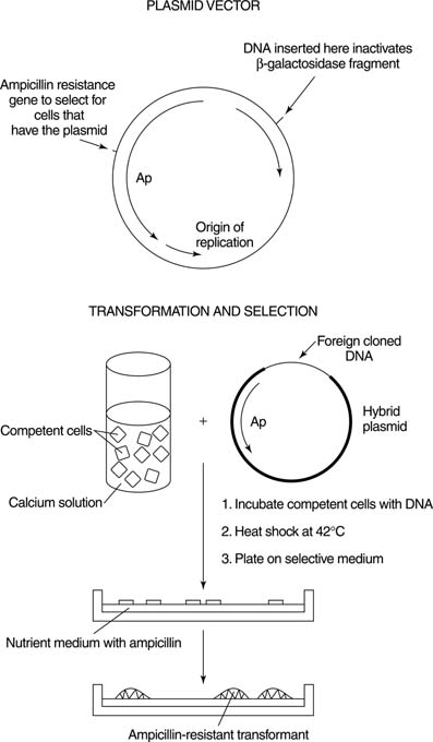
Plasmids are circular DNAs that are capable of independent replication. Many naturally occurring bacteria contain plasmids; plasmid vectors are derived from naturally occurring plasmids. Plasmid vectors have several properties. First, they contain single restriction sites for several enzymes. Cleaving with one of these enzymes generates a single, linear form of the plasmid. This feature helps to ensure efficient ligation, because every ligation product will contain the entire vector sequence. Secondly, vectors are made to contain selectable genetic markers so that cells which contain the vector can be propagated. For example, many plasmid-cloning vectors contain a gene that encodes resistance to the antibiotic ampicillin (a member of the penicillin family). Cells that contain the vector can then be selected for by growth in the presence of the antibiotic; cells that don't contain the vector sequence will be inhibited by ampicillin in the growth medium, as shown in Figure 1 . 


Figure 1

Thirdly, a means to detect which cells have only the plasmid vector as opposed to the recombinant product must exist. This determination is usually accomplished by a mechanism called insertional inactivation. The idea of insertional inactivation is that inserting passenger DNA into a gene interrupts the sequence of the gene, thereby inactivating it. For example, the restriction sites of many common plasmid-cloning vectors are located in a fragment of a gene for β-galactosidase, an enzyme involved in lactose metabolism. When cells containing just the vector are grown in the presence of an artificial substrate related to lactose, the colonies turn blue, because active enzyme is made. On the other hand, when the restriction site has a piece of foreign DNA inserted into it, the gene cannot make an active protein fragment because the DNA sequence interrupts the coding sequence of the gene. As a result, colonies of the bacteria that contain cloned foreign DNA appear whitish. The bacteria that are present in the colony can be grown separately, and standard biochemical procedures easily isolate the recombinant DNA they contain. If no such selection existed, each colony would have to be grown separately and its DNA analyzed—a very “hit or miss” proposition in many cases.
After a recombinant plasmid has been formed in the laboratory, it must be replicated. This process begins with the growth of the cell containing the recombinant plasmid. Plasmids are usually transferred to new hosts bytransformation. Transformation is the addition of naked foreign DNA into a recipient bacterium. Because the plasmid usually contains a gene encoding the ability to grow in the presence of an antibiotic, identifying a recombinant bacterium is a two-part process. First, growth in the presence of the antibiotic ensures that each bacterial colony contains a plasmid, while the color of the colony identifies the plasmids that contain inserted DNA. Finally, the growth of the transformed bacterial colony amplifies the clones, that is, makes more copies of the plasmid. These steps can be done with a known piece of DNA or with a less-defined collection of insert DNA— for example, all the DNA from an organism. A collection of different cloned DNAs is called a library. The number of independent sequences in a library is called its complexity; the more complex the library, the greater number of independent sequences it contains.

Other types of cloning vectors

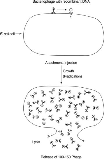
Viruses that infect bacteria are called bacteriophages. Native bacteriophage have been formed into vectors that can also be used for cloning. Bacteriophage vectors have three advantages over plasmids. First, they can carry significantly more foreign DNA than can plasmids, which are limited to about 5,000 base pairs (5 kilobases) of foreign DNA. Plasmids with larger-sized inserts tend to be unstable when amplified. The larger the insert, the fewer independent clones are required to have a reasonable chance of identifying any single gene or sequence in the collection of cloned DNA. Secondly, the virus particles of bacteriophage vectors can accept DNA only of a narrow size range. This means that DNA can be preselected so that each recombinant virus will contain only a single foreign sequence. This property is a consequence of the fact that DNA (whether native or foreign) must be packaged into a protein coat to be infectious. DNAs that are too large or too small can't be packaged efficiently, so they will not be represented in the library. Finally, bacteriophage infection can be a very efficient process, with nearly 100 percent of the packaged virus particles leading to a productive infection. In contrast, cells take up only one of 100,000 plasmid molecules in a transformation procedure. Bacteriophage clones are amplified by repeated cycles of infection and growth. See Figure 2 . 


Figure 2

The Polymerase Chain Reaction


You can isolate virtually any DNA sequence by means of the polymerase chain reaction, or PCR. PCR uses repetitive cycles of primer-dependent polymerization to amplify a given DNA. Very little original DNA is required, as long as two unique primers are available. Knowing the sequences of the primers before starting out is helpful, but not always necessary. Each cycle of PCR involves three steps: DNA double strand separation, primer hybridization, and copying. First, the original DNA is denatured by heat treatment to make two separated strands. Then the two primers are hybridized to the DNA, one to each of the two separated strands. These primers act as initiators for DNA polymerase, which copies each strand of the original double-stranded DNA. The original two strands of DNA now become four strands, which are then denatured. These four strands are then hybridized with the primers and each of them is now copied, to make eight strands, and so forth. Amplified DNA can be analyzed by any of the techniques used for analyzing DNA: it can be separated by electrophoresis, Southern blotted, or cloned. Because a single DNA sequence is obtained by PCR, sequence information can also be obtained directly. See Figure 1 . 


Figure 1

DNA Sequence Determination


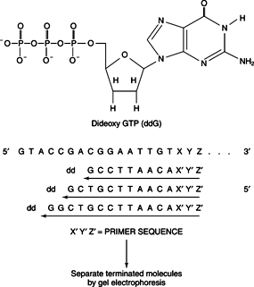
The most important information about DNA is its nucleotide sequence. DNA sequences are determined by exploiting the enzymatic characteristics of DNA polymerase. DNA polymerases all require a primer to initiate synthesis. The sequencing reaction begins when a single primer is hybridized to the strand of DNA to be sequenced. This primer-initiated synthesis is just like the initiation of a PCR. The primer used to initiate synthesis can be complementary to the vector in which the DNA is cloned, or it can be a primer used to amplify the DNA in PCR. The important point for sequencing DNA is that all of the newly synthesized molecules will have the same sequence. The primer is extended by addition of deoxynucleoside triphosphates along with DNA polymerase. If these were the only components of the reaction mixture, then all the newly extended molecules would be complementary to the entire template chain, just as in PCR. The sequencing reaction also contains, in addition to the four deoxynucleoside triphosphates (dNTPs), 2′,3′-dideoxynucleoside triphosphates, or ddNTPs (ddGTP, ddATP, ddTTP, or ddCTP), as shown in Figure 1 . 


Figure 1

As the preceding figure shows, the ddNTPs lack both the 2′ and 3′ hydroxyl groups of a ribonucleoside triphosphate; therefore, if a ddNTP is incorporated into the growing DNA chain, all synthesis stops. Because a ddNTP must be base-paired for it to be inserted into a chain, the incorporation of a dideoxynucleotide at a position is an indicator of the nature of the complementary base on the chain. In other words, ddG is incorporated into the chain only when a C is present in the template, ddA only in response to a T, and so on. Because either a deoxy- or dideoxynucleotide may be inserted at a given site, the result is a set of nested chains, beginning at the primer and ending at a dideoxynucleotide. The members of this collection of molecules all have the same 5′ end (because they all start with the same primer) and their 3′ ends terminate with a dideoxynucleoside. If the molecules are separated according to chain length by electrophoresis through a thin polyacrylamide gel, then the nucleotide sequence of the corresponding DNA can be read directly. The DNA fragments are detected by fluorescence. Sequences are read automatically from a single lane because each terminator is modified to contain a different fluorescent dye, so that each dideoxynucleoside is associated with a single color of the fluorescence. By using these methods, it is possible to read 700 or more nucleotides in a single experiment.

Genomics


With easy DNA sequencing technology comes the possibility to determine all the information present in the DNA of a single organism—the entire sequence of its DNA. Genomics is the study of an organism's entire information content. In humans, that amounts to about 3 billion base pairs of information, or about 10 million sequencing experiments (although it isn't nearly all that efficient). More to the point is the sequence information in a small bacterium, about 4 to 5 million base pairs of information. This is much more feasible, and can be accomplished in about 10,000 to 20,000 separate experiments—a few weeks' worth of information gathering in a big, dedicated laboratory. The strategy is sometimes called “shotgun sequencing,” because the information-gathering process isn't precisely aimed. Rather, a random set of clones is made in a bacteriophage-like vector (called acosmid) and a mini-library of DNA inserts is made in a plasmid vector. These recombinant plasmids are then used as templates for sequencing reactions such as the ones described earlier. The sequences are analyzed by computer so that the overlapping DNA sequences can be fit together to make longer sequences. This step-by-step process is continued until enough information is gathered to assemble a whole sequence of the DNA for an organism or chromosome. Although the shotgun technique may lead to a single stretch of sequence being determined several times before all the sequences are determined, it is faster than trying to order each clone and then determine their sequences individually.
Scientists have determined complete genomic sequences for a number of viruses, over fifteen bacteria, and laboratory yeast. Many pharmaceutical companies are interested in the sequence information from bacteria that cause disease, in the hopes that the information obtained will lead to new drug targets.

DNA Hybridization


Nucleic acid hybridization allows scientists to compare and analyze DNA and RNA molecules of identical or related sequences. In a hybridization experiment, the experimenter allows DNA or RNA strands to form Watson-Crick base pairs. Sequences that are closely related form base-paired double helices readily; they are said to be complementary. The amount of sequence complementarity is a measure of how closely the information of two nucleic acids relate. The complementary strands can be both DNAs, both RNAs, or one of each.
Heating the DNA solution above a characteristic temperature can separate the two strands of a double helix. That temperature is called the melting temperature, abbreviated Tm . Above the Tm, a DNA is mostly or all single-stranded; below the Tm, it is mostly double-stranded. For a natural DNA, the Tm depends primarily on its G+C content. Because a G–C base pair has three hydrogen bonds and an A–T pair only has two, nucleic acid double helices with a high G+C content have a higher Tm than do those with a greater proportion of A+T. The Tm is not an exact property: It depends on the solvent conditions. For example, a high concentration of salt (such as NaCl) raises the Tm of a DNA duplex, because the positive Na+ ions shield the negative charges on the phosphodiester backbone from repelling each other. Likewise, certain organic solvents can cause the negative charges on the phosphates to repel more strongly; these solvents lower the Tm of a DNA double helix.
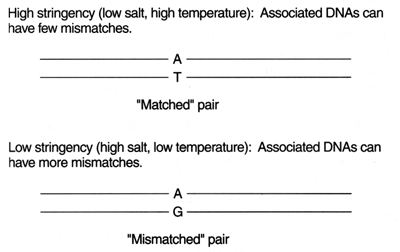
What happens if two nucleic acids are partly complementary and partly different? In this case, some stretches of the two strands may form base pairs while others don't. The two molecules can be manipulated so that they form a hybrid or separate. The conditions favoring the formation of duplex nucleic acid are low temperature (below the Tm), high salt, and the absence of organic solvents. The latter two conditions raise the Tm of the hybrid duplex so that the DNA would remain more double-stranded. On the other hand, higher temperatures (closer to the Tm of the hybrid) lower salt, and the presence of organic solvents would tend to push the two strands of the DNA apart. The term stringency sums up these variables: The more stringent the conditions, the more likely partially complementary sequences are to be forced apart. Conversely, less stringent hybridization conditions mean that the two strands need not be so complementary to form a stable helix. See Figure 1 . 


Figure 1

Hybridization can be used to classify the DNAs of various organisms. For example, human DNA is 98 percent identical to that of chimpanzees, and these two DNAs form a duplex under stringent conditions. Related sequences of humans and birds can also form hybrids, but only at a much lower stringency.